Serving Your Vision
Slit Lamp Microscope
Slit Lamp Microscope
| Model Name | Manufacturer |
|---|---|
| 700GL NSW Slit Lamp Microscope | Takagi Seiko Co., Ltd. |
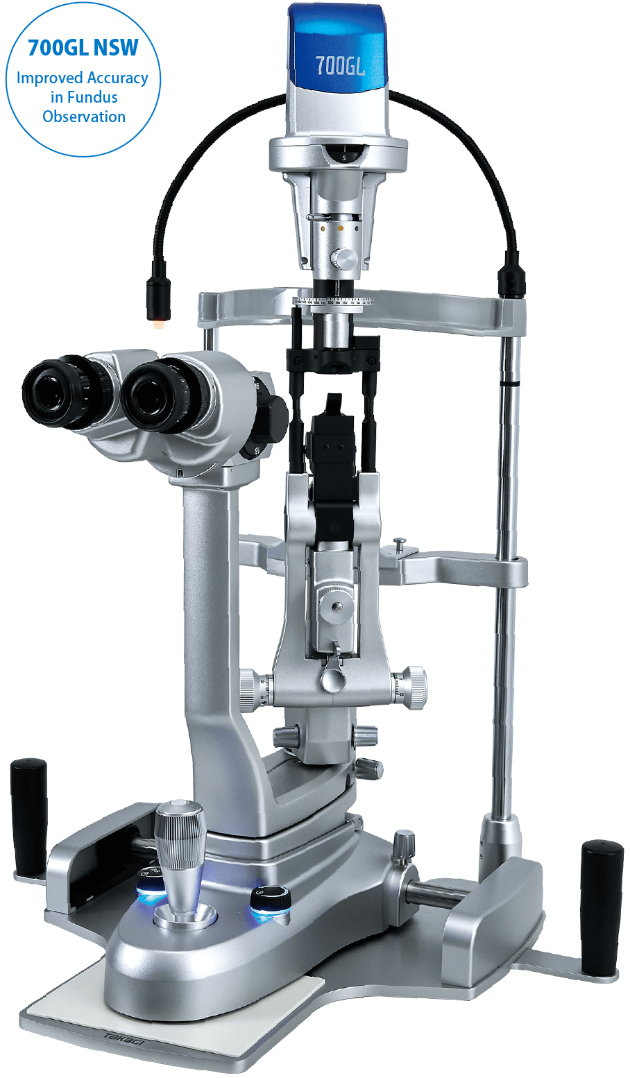
A slit lamp is an essential instrument for the diagnosis of various eye diseases.
The 700GL NSW is equipped with innovative NSW function to enable observations in even greater detail.
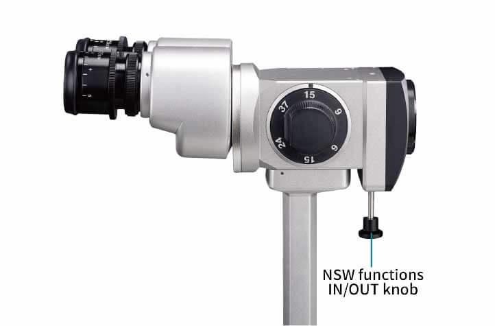
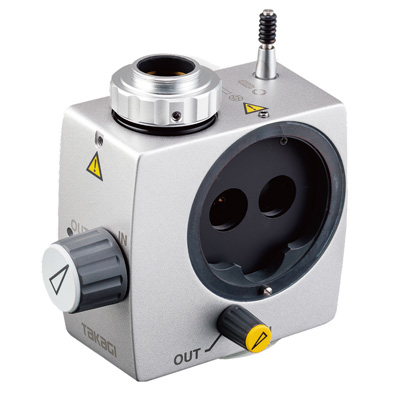
In contrast to the 10° stereo angle of the 700GL, an angle of 4.1° is adopted for the NSW, ensuring a wider stereoscopic vision area. This makes it easier to observe the fundus in detail for highly accurate diagnosis. *700GL NSW is a factory option specification. Please note that the standard 700GL cannot be modified/converted to 700GL NSW after purchase. NSW is an acronym of “Narrow angle,” “Stereo,” and “Wide viewer.”
*The image is for illustrative purposes only.
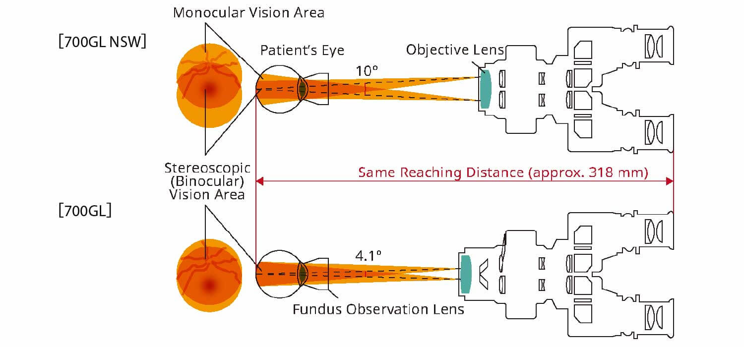
*The image is for illustrative purposes only.
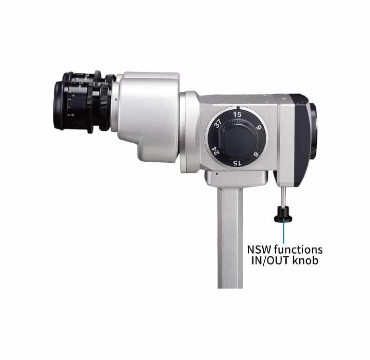
Switching to the NSW functions is easy. Simply press the knob at the bottom of the objective lens.
The inter-optical path distance of 22 mm and the sharp slit illumination that uses a high-luminance LED with a 3500 K colour temperature produce a clear observation image with a stereoscopic feel from the anterior eye parts to the fundus. Lesions and foreign matter can be identified clearly and in detail, supporting more accurate diagnosis.

The inter-optical path distance of 22 mm and the sharp slit illumination that uses a high-luminance LED with a 3500 K colour temperature

*Photo provided by: Dr. Toru Noda, Department of Ophthalmology, NHO Tokyo Medical Center
To reduce the burden on doctors during long examinations and minimise fatigue, the design focuses on ease of use, from the size and positioning of the operating parts to their operational feel. This model uses an energy-saving LED light source which provides stable brightness over a long service life. This allows a design with built-in cables and reduces heat generation, contributing to enhanced patient safety.

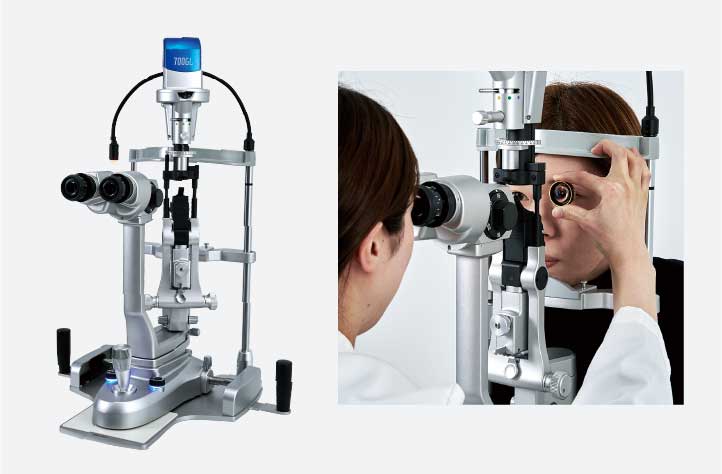
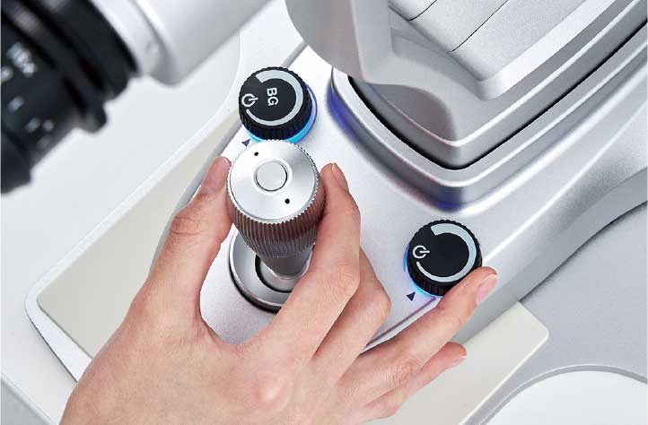
The design consists of parts such as a large-diameter dimmer knob arranged functionally around the joystick for easy operation with one hand.

The size and feel of the large-diameter control knob are optimally designed for comfortable operation.

The front lens can be held in a stable position using the finger rest function of the forehead rest. This makes observations easier and reduces the burden on the doctor’s arms, while also minimizing contact with patients.

Background illumination in use
The background illumination uses an LED with the same colour temperature as the slit illumination to obtain a clear image without impairing the colour balance.
This makes the entire eye clearly visible, enabling patients to intuitively understand the location and condition of the affected area.

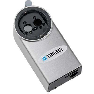
A camera can be connected for imaging by attaching a TAKAGI combination or camera adapter. A TAKAGI digital camera is also available.

Simply pressing a button once to capture an image while operating the joystick.
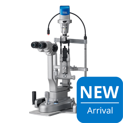
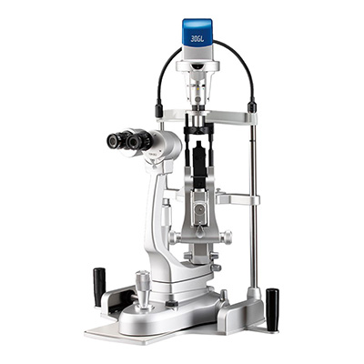
3-step Galilean type, converging optics, upper LED illumination, without background illumination
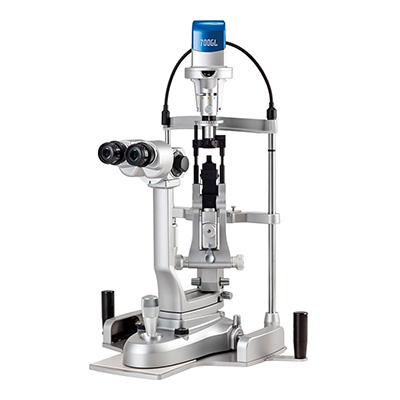
5-step Galilean type, converging optics, upper LED illumination, and built-in background illumination
5-step Galilean type, converging optics, upper LED illumination, built in background illumination and wide angle fundus observation
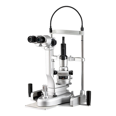
2-step Greenough type with converging optics and upper LED illumination
5-step Galilean type, converging optics, lower LED illumination, and built in background illumination
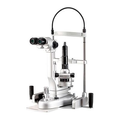
3-step Galilean type with converging optics and lower LED illumination

Portable, LED lighting, anterior segment observation, excellent operability
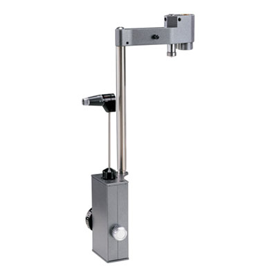
R-Type Compact Applanation Tonometer with option to be permanently fixed to slit lamp with observation from the left eye
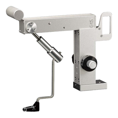
Z-type applanation tonometer exclusively for use with 2ZL/4ZL
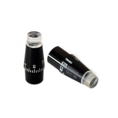
Reusable Tonometer Measuring Prism
 Home
Home Distributors Login